What are the main components of the circulatory system?
The circulatory system has three main components:
- blood
- blood vessels
- the heart
It has two main functions:
- transportation of substances
- protection against disease
Parts of the blood
The main blood components are:
| Component | Function | Structure |
|---|---|---|
| Red blood cell | Carry oxygen | Contain haemoglobin (rich in iron) to carry oxygen. Biconcave shape to provide a large surface area for the diffusion of oxygen. No nucleus to provide more space for haemoglobin |
| White blood cell | Defend the body against disease | Large cells that contain a nucleus. There are two types: lymphocytes – make antibodies. Phagocytes – engulf and digest microorganisms |
| Platelets | Convert fibrinogen to fibrin. Fibrin forms a mesh that traps blood. Important in blood clotting and scab formation | Cell fragments (very small) |
| Plasma | Transports blood cells, digested food molecules (eg glucose), carbon dioxide, urea and hormones | Yellow coloured liquid |
What is the role of platelets in blood clotting and scab formation?
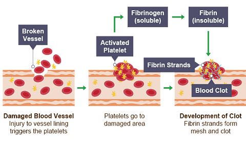
- Blood vessel is damaged
- Platelets triggered
- Convert fibrinogen (soluble) into fibrin (insoluble)
- Forms a mesh or net
- Traps the blood cells
- A clot is formed
- Dries to form a scab
Cell lysis
If red blood cells are placed in a high concentration of water, water will move into the cell by The process by which water moves in and out of cells from a high concentration to a low concentration. and they will lyse (burst) because they don’t have a cell wall.
The blood vessels
Structure
| Feature of blood vessel | Artery | Vein | Capillary |
|---|---|---|---|
| Wall thickness | thick | thin | very thin - one cell thick |
| Muscle and elastic fibres | thick layer | thin layer | none |
| Lumen diameter | small | large | very small - one cell thick |
| Valves | no | yes | no |
Arteries
Carry Blood that is high in oxygen and low in carbon dioxide. blood away from the heart to the body (except the pulmonary artery, which carries Blood that is low in oxygen (as cells have used it) and high in carbon dioxide (as cells have produced it). blood to the lungs).
Thick walls with muscle and elastic fibres to withstand high blood pressure and allow expansion and recoil with each heartbeat.
Veins
Carry deoxygenated blood back to the heart (except the One of the four veins that carries oxygenated blood to the heart from the lungs., which carries oxygenated blood from the lungs).
Blood is moving at a low pressure, so the walls are thin.
The structure in veins that prevents the backflow of blood. prevent backflow of blood.
Capillaries
Allow exchange of substances (eg oxygen, carbon dioxide, food, urea) between blood and cells.
Walls are If a substance is permeable, it allows liquids or gases to go through it. and one cell thick providing a short diffusion path.
The heart
The heart is a unidirectional pump.
Valves prevent the backflow of blood ensuring unidirectional flow.
The right-side pumps Blood that is low in oxygen (as cells have used it) and high in carbon dioxide (as cells have produced it). blood to the lungs.
The left side pumps Blood that is high in oxygen and low in carbon dioxide. blood to the organs of the body.
The heart has four chambers - right atrium, right ventricle, left atrium, left ventricle.
The left ventricle wall is thicker than the right ventricle because it pumps blood around the body while the right ventricle pumps blood to the lungs, located close to the heart.
Coronary arteries supply the heart muscle with oxygen and glucose for respiration. They can be seen on the external surface of the heart.
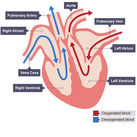
The heart in one heartbeat
Deoxygenated blood enters the right atrium from the vena cava
Blood moves into the right The lower chambers of the heart which receive blood from the atria and pump it to the body. The right ventricle pumps it to the lungs and the left ventricle pumps it to the rest of the body.
Deoxygenated blood is pumped into the pulmonary artery to the lungs
Blood becomes oxygenated in the lungs
Oxygenated blood leaves the lungs via the pulmonary vein
Blood enters the left atrium
Blood moves into the left The lower chambers of the heart which receive blood from the atria and pump it to the body. The right ventricle pumps it to the lungs and the left ventricle pumps it to the rest of the body.
Blood is pumped into the aorta to circulate oxygenated blood around the body
Double circulatory system
Mammals have a double circulatory system, where blood passes through the heart twice in one full circulation.
The following arteries and veins transport blood to and from some of the body’s organs:
| Blood vessel | Function |
|---|---|
| Vena cava | carries deoxygenated blood from the body back to the heart |
| Pulmonary artery | carries deoxygenated blood from the heart to the lungs |
| Pulmonary vein | carries oxygenated blood from the lungs to the heart |
| Aorta | carries oxygenated blood from the heart around the body |
| Hepatic artery | carries oxygenated blood to the liver |
| Hepatic vein | carries deoxygenated blood back to the heart. Carries digested food (glucose and amino acids) from the liver around the body |
| Hepatic portal vein | carries digested food from the small intestine to the liver |
| Renal artery | carries oxygenated blood (also rich in urea) to the kidneys for excretion |
| Renal vein | carries deoxygenated blood (also low in urea as it has been purified in the kidney) back to the heart |
Why is exercise so beneficial?
During exercise, muscles contract more and require more energy, which is released through The chemical change that takes place inside living cells, which uses glucose and oxygen to release the energy that organisms need to live. Carbon dioxide is a by-product of respiration..
Respiration needs glucose and oxygen which is carried in the blood to the muscles.
To meet this demand, the blood flow to the muscles increases, raising the pulse rate (heart rate).

Regular exercise is beneficial as it:
strengthens the heart muscle
increases cardiac output when at rest – lower pulse rate
reduces chance of a heart attack
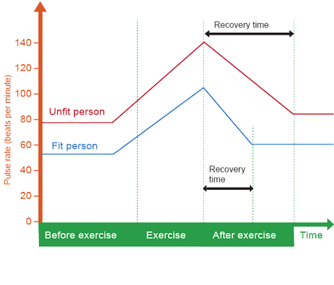
It also reduces the recovery rate (time taken for heart rate to return to normal after exercise).
Watch: How our circulatory system keeps us alive
How much do you know about the circulatory system?
More on Body systems
Find out more by working through a topic
- count3 of 3

- count1 of 3